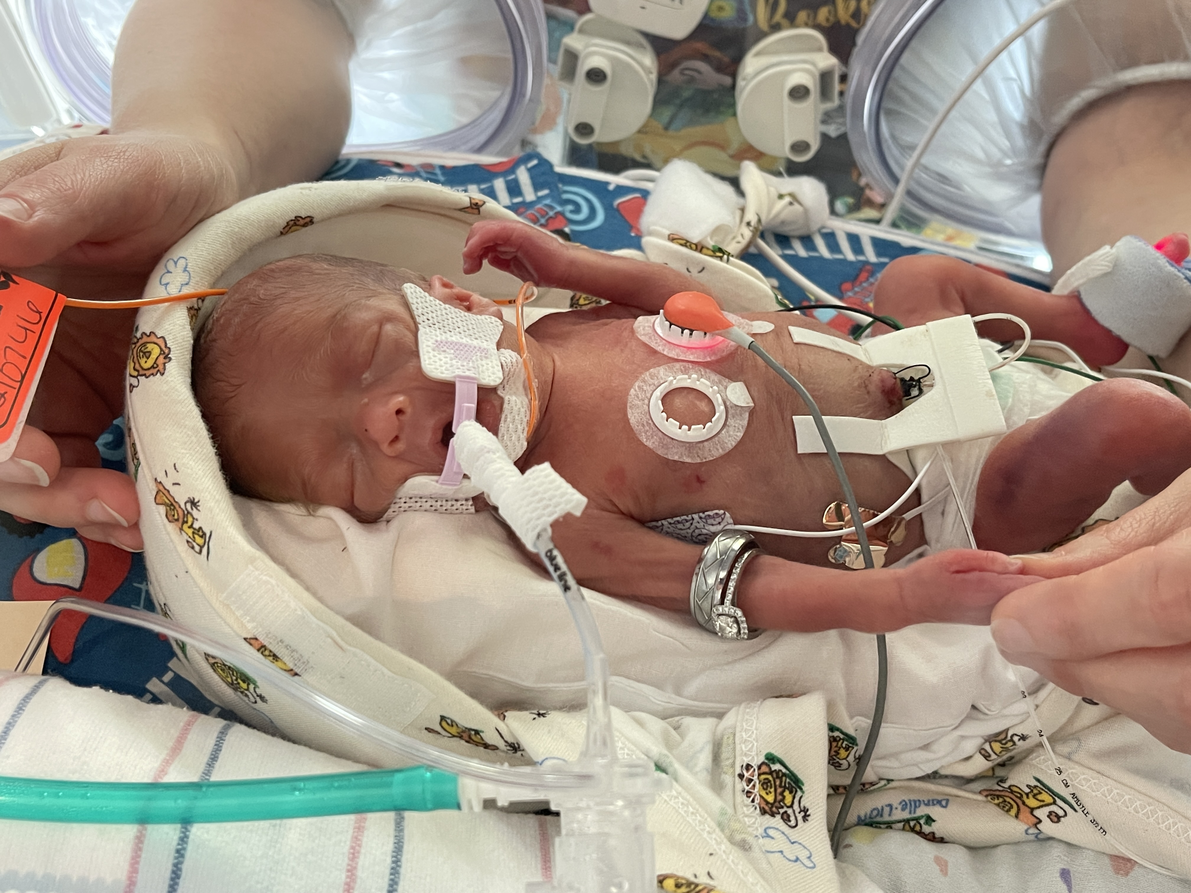
In the NICU, closely monitoring carbon dioxide (CO2) levels is a crucial aspect of care for premature and critically ill infants — fluctuations in CO2 levels can disrupt cerebral blood flow, increasing the risk of intraventricular hemorrhage (IVH), periventricular leukomalacia (PVL), and other cerebral injuries.¹ Continuous visibility of CO2 can help care teams to carefully target levels within a safe range, helping to avoid these dangerous complications.
End-tidal carbon dioxide (etCO2) monitoring, also known as capnography, is one method that can be used to continuously monitor CO2. This technique measures the concentration of CO2 in exhaled breath and is a particularly useful tool for quickly confirming proper endotracheal tube (ETT) placement — often more effectively than standard clinical assessments.¹ Additionally, it provides a waveform that clinicians can interpret for insights into airway resistance and lung compliance. However, despite these benefits, etCO2 monitoring has limitations specific to neonatal patients that NICU teams must keep in mind when choosing the most appropriate monitoring method.
Limitations of End-Tidal CO2 Monitoring in the NICU
 Endotracheal tube leaks can make etCO2 inaccurate
One of the main challenges with end-tidal CO2 monitoring occurs when using an uncuffed endotracheal tube (ETT). In neonates, uncuffed ETTs are usually preferred to reduce the risk of airway damage, post-extubation stridor, and other complications associated with intubation. However, air leakage around the tube is common, affecting approximately 70% of ventilated neonates.² This leakage can cause the gas sample measured by an end-tidal monitor to inaccurately represent the patient’s actual PaCO2 levels, as some exhaled gas escapes around the tube and bypasses the sensor.³ This issue is concerning when managing intubated neonates, as precise monitoring is critical for adjusting ventilatory settings and ensuring effective gas exchange.
 EtCO2 adds dead space to the circuit and weight to the ET tube
Another challenge with end-tidal CO2 monitoring is that the additional volume from etCO2 monitoring equipment, such as sensors and sampling lines, increases dead space — the portion of each tidal volume that does not participate in gas exchange. The addition of dead space during end-tidal CO2 monitoring can cause rebreathing of exhaled CO2, leading to inaccurate expiratory measurements. 4 This issue is especially problematic in preterm infants, where even a small addition of dead space can have a large impact on their dead space to tidal volume ratio (Vd/Vt), further complicating the accuracy of readings.
The added weight of end-tidal CO₂ monitoring equipment on a neonate’s ETT is another challenge. While many newer etCO2 devices are designed to be lightweight, even a small amount of additional weight is an important consideration, as the ETT itself already presents a risk of trauma to the delicate airway.
 EtCO2 is less accurate with small tidal volumes and high respiratory rates
While low tidal volumes and high respiratory rates are often optimal for preterm infants to protect their delicate lungs, together they introduce technical challenges when attempting to monitor CO2 using an end-tidal device. For most premature infants, the target tidal volume ranges from 4-6 ml/kg, so for the smallest patients, it can be as low as 2-3 ml. When monitoring CO2 through an end-tidal device, a small tidal volume becomes a challenge due to the added dead space previously mentioned. The dead space volume in many end-tidal CO2 devices is around 1 mL, which may seem small, but for neonates with these low tidal volumes, it can become a significant portion of the total exhaled volume. 4
Furthermore, a high respiratory rate exacerbates the inaccuracy of etCO2 readings in these infants. Preterm infants typically have respiratory rates of 40-60 breaths per minute, with critically ill neonates often exceeding that. 4 These infants have a short expiratory time, which also presents technical limitations for end-tidal monitoring due to the devices limited response time — the shorter the expiratory time, the shorter the measurement window.
These challenges are especially pronounced in infants receiving high-frequency ventilation, such as high-frequency jet ventilation (HFJV) and high-frequency oscillatory ventilation (HFOV). These specialized modes deliver extremely small tidal volumes at very high respiratory rates, often between 200-900 breaths per minute. In such cases, end-tidal monitoring becomes impractical, as it cannot accurately capture each breath through the sensor.
 EtCO2 is known to be unreliable with ventilation/perfusion (V/Q) mismatch
V/Q mismatch happens when either a patient’s ventilation (V) or perfusion (Q) is impaired, hindering gas exchange at the alveolar level. This can occur in neonates with bronchopulmonary dysplasia (BPD), meconium aspiration syndrome (MAS), respiratory distress syndrome (RDS), and other conditions. Whether the patient’s issue is related to ventilation, perfusion, or both, a V/Q mismatch leads to discrepancies between the CO2 levels in exhaled breath and those in the blood. As a result, an end-tidal measurement commonly underestimates these patients’ actual PaCO2 values — in some cases significantly so. 5
The inability to effectively monitor CO2 with end-tidal in these cases poses a significant limitation for care teams, as accurate CO2 monitoring is important for ensuring optimal management while neonates (or patients of any age) are on respiratory support.
 EtCO2 is incompatible with noninvasive ventilation (NIV) modalities like NPPV, HFNC, NIV-NAVA, and bubble CPAP
Noninvasive ventilation (NIV) modalities are increasingly chosen in the NICU to protect delicate neonatal lungs. Noninvasive methods like continuous positive airway pressure (CPAP), high-flow nasal cannula (HFNC), and noninvasive positive pressure support (NIPPV) provide respiratory support without the need for intubation, reducing the risk of airway damage and other complications associated with invasive mechanical ventilation. However, etCO2 monitoring is generally incompatible with noninvasive or specialty ventilation modalities, limiting its utility in clinical situations where these strategies are preferred.
Why Not Just Draw a Blood Gas?
Given the importance of closely monitoring a neonate’s CO2 levels, blood gas tests are routinely used in the NICU, particularly in cases where end-tidal CO2 measurements are unreliable. While blood gas measurements are undoubtedly an important part of care in the NICU, they can be a major source of both pain and blood loss for these tiny patients. Iatrogenic blood loss, anemia of prematurity, and risks associated with transfusions should be taken into consideration. In addition, mounting research shows that repeated exposure to pain and stress in the NICU can have a negative impact on neurological development.
This presents a challenge: How can NICUs watch and manage CO2 levels closely, accurately, and continuously, especially as patients transition between different forms of respiratory support?
Considering Transcutaneous CO2 Monitoring for the NICU
Understanding the limitations of end-tidal monitoring is important for NICU care teams as they optimize care for their patients. End-tidal CO2 monitoring can be valuable in certain clinical contexts, providing real-time breath-to-breath waveforms and facilitating the placement of ETTs. Nonetheless, inherent technological limitations of this tool can complicate the accurate assessment of a patient’s respiratory status.
For NICU teams aiming to address these limitations, transcutaneous CO2 monitoring is a valuable tool worth considering. This technology works by continuously and noninvasively measuring the diffusion of CO2 through the skin and using that measurement to approximate PaCO2. This measurement can help teams guide effective care for even the most fragile patients.
Unlike etCO2, transcutaneous monitoring is compatible with any mode of ventilation — including mechanical ventilation, high-frequency ventilation (HFV), high-flow nasal cannula (HFNC), and bubble CPAP. This versatility allows it to be used across a wide range of clinical scenarios, providing accurate, real-time CO2 data without being restricted by the type of respiratory support.
Furthermore, transcutaneous monitoring does not introduce dead space or added weight on the ETT, as it uses a sensor applied to the skin rather than equipment attached to the airway. Transcutaneous monitoring maintains accuracy regardless of ventilation-perfusion (V/Q) mismatch, variations in respiratory rate or tidal volume, supporting reliable monitoring of gas exchange across a variety of clinical conditions.
For Your Team: The NICU Pocket Guide
Transcutaneous CO2 monitoring can be a valuable tool for your small baby unit care. Our Clinical Pocket Guide on Transcutaneous CO2 Monitoring for Neonates can assist your team in integrating this technology into your unit’s protocols or enhancing its use to support patient care.
Let’s talk!
Interested in exploring transcutaneous CO2 monitoring for your NICU?
We would love to learn about your unit and discuss how our transcutaneous monitoring system can enhance your patient care. You can also request a hands-on demo with your team so you can experience the technology for yourselves.
References
- Hochwald, O., et al. Continuous Noninvasive Carbon Dioxide Monitoring in Neonates: From Theory to Standard of Care. Pediatrics. 2019.
- Bernstein, G., et al. Airway leak size in neonates and autocycling of three flow-triggered ventilators Crit Care Med. 1995.
- Takahashi, D., et al. Effect of tidal volume and end tracheal tube leakage on end-tidal CO2 in very low birth weight infants. J Perinatol. 2021.
- Schmalisch, G. Current methodological and technical limitations of time and volumetric capnography in newborns. Biomed Eng Online. 2016.
- Cox, P., et al. Noninvasive Monitoring of PaCO2 during One-Lung Ventilation and Minimal Access Surgery in Adults: End-Tidal versus Transcutaneous Techniques. J Minim Access Surg. 2007.


